- Research studies
The Department of Optometry and Vision Sciences is committed to training and supporting our next generation of vision researchers, no matter what your major there are vision research pathways for you.
- Alumni
Our extensive network of alumni and friends include local and international graduates who have followed diverse career paths from research to primary care, industry to politics.
Department of Optometry and Vision Sciences - Research
Learn more about honours, masters, and graduate research
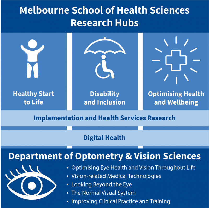
Research led by the Department of Optometry and Vision Sciences sits primarily within the Optimising Health and Wellbeing hub. The majority of our work is designed to improve understanding and outcomes for a range of eye and vision disorders. These also include brain diseases that manifest in the eye and the development of new technologies, diagnostics and therapeutics to overcome them.
Our researchers in the Healthy Start to Life hub focus on vision development in children. In the Disability and Inclusion hub, our researchers focus on improving outcomes and developing medical technologies for people with vision loss, visual function disorders, ocular and brain disease as well as methods to optimise delivery of patient care.
Underpinning research in these hubs is research in Implementation Science and Digital Health, including investigating new approaches to enhance optometric training and improve clinical practice.
The Department of Optometry and Vision Sciences’ Graduate Research and PhD programs strive to advance eye care and vision, within Australia and internationally, through excellence in research spanning bench to bedside.
Explore our programs of research
-
Optimising Eye Health and Vision Throughout Life
This program of work focuses on fundamental and applied research on vision development and visual pathways, and studying how they can be altered with ageing, injury and disease. Our research includes the development of methods to enhance the detection and monitoring of eye disease, and investigating novel treatments and management options, including vision restoration. This program incorporates research into the prevention of eye disease, improving health services and access to clinical care.
-
Vision-related Medical Technologies
This program of work focuses on investigating novel medical and ocular technologies for the detection and management of ocular disease and systemic conditions that affect the eye. Our research also includes multidisciplinary and collaborative interactions with other academic departments and industry to develop, evaluate and optimise diagnostic tests and therapeutics including novel ways to study the eye and brain. The bench-to-bedside research program spans many levels of the visual system and all stages of technology and drug development, including clinical trials and implementation studies.
-
Looking Beyond the Eye
This program focuses on research into conditions that can affect the eye, visual pathways and the brain. Investigating the eye can provide valuable insight into a range of systemic conditions, such as vascular and cortical disease. In addition, the brain can provide valuable insight to a wide-range of perceptual disorders. Fundamental and applied research streams focus on investigating the interplay between vision, the eye and the brain, in ageing, injury and disease.
-
The Normal Visual System
This program of research focuses on investigating the normal visual system and how it performs in different environments. We aim to further discovery in how the eye and visual pathways work. Fundamental and applied research streams focus on using the visual system to provide insight into the integrative functions of the brain such as attention, perceptual learning and decision making and the neural circuits underpinning them.
-
Improving Clinical Practice and Training
This program of research focuses on developing, implementing and evaluating evidence-based strategies to improve and optimise eye care practice and training, patient care and service delivery. Harnessing digital and learning technologies, we develop teaching initiatives to improve optometric education and undertake rigorous evidence syntheses to support evidence-based practice.

Melbourne Eyecare Clinic
Melbourne Eyecare Clinic is a key collaborator with the Department in clinical research. The clinic provides a teaching environment for optometry students at the University of Melbourne and delivers an extensive range of eyecare services to the public. The clinic has a comprehensive array of state-of-the-art diagnostic imaging equipment and other specialist tools, enhancing clinical research capabilities available at the Department.
The School educates graduate entry and post-graduate students through accredited programs tailored to workforce needs nationally and internationally, enabling them to be competent and effective health professionals who are work ready and eligible for registration.
-
Degrees
Our graduate entry and post-graduate degrees are tailored to workforce needs nationally and internationally.
-
Honours in Vision Sciences
Look deeper into the Science of Vision as part of your Honours programme at the University of Melbourne. Research projects for Honours (Vision Science) can be viewed here.
-
Scholarships, Bursaries and Prizes
The Faculty of Medicine, Dentistry and Health Sciences offer an extensive range of scholarships and bursaries to undergraduate and postgraduate coursework students.
-
Facilities & Resources
The Department has access to a number of facilities and provides a number of resources for its students, staff and supervisors.

Student Placements
Work-integrated learning, including placement, provides career-defining experiences for students and is integral to many programs within
the Faculty of Medicine, Dentistry and Health Sciences.
The Department's research and teaching success is underpinned by active engagement with key stakeholders, the community and the profession.
-
Alumni
The department has a focus on engagement and interaction with the community. We hope to keep all colleagues who graduated from the Department in touch with University of Melbourne news and views.
-
Giving
We are grateful for the many ways in which our alumni and donors support the work of the Faculty.
-
Partners
The Department has new relationships and long-standing affiliations with a number of organisations.
-
Community
A key focus of the Department's engagement strategy has been the increase in the connection to the community and industry through the Melbourne Eyecare Clinic.

Welcome to the Department of Optometry and Vision Sciences
Associate Professor Andrew Metha
The Department of Optometry and Vision Sciences educates future optometrists, performs internationally recognised vision science research, and contributes widely to the advancement of optometry as an essential health care discipline.
Melbourne Eyecare Clinic
The Department of Optometry & Vision Sciences operates the Melbourne Eyecare Clinic which offers patient care primarily for University staff and students, but is also open to the general public and for specialist referral by other practitioners.